The humble bird’s egg represents one of nature’s most perfect designs—a self-contained life support system that transforms seemingly simple contents into a fully-formed chick ready to face the world. While we may admire eggs from the outside, the miraculous journey happening within remains largely unseen. From a single fertilised cell to a complex living creature ready to crack through its shell, the development of a bird embryo is a fascinating process governed by precise biological timing and genetic programming. As the developing chick grows within its calcium carbonate fortress, it undergoes remarkable transformations, forming organs, limbs, and specialised adaptations needed for survival. Let’s crack open the science behind this incredible developmental journey and explore the hidden world inside a bird’s egg before hatching day arrives.
The Foundation: Egg Structure and Components
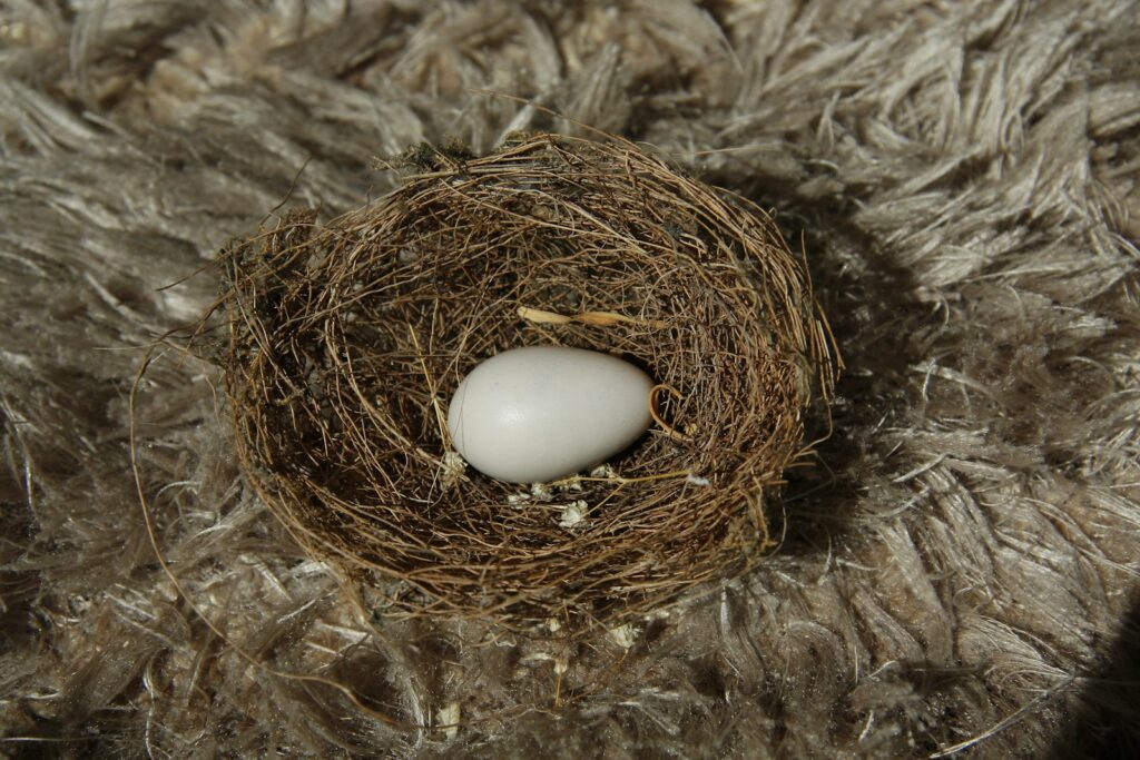
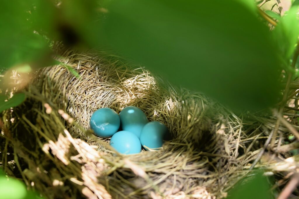
A bird’s egg is far more complex than it appears, with specialised structures that support embryonic development. The hard outer shell, composed mainly of calcium carbonate, provides physical protection while remaining porous enough to allow oxygen and carbon dioxide exchange. Beneath the shell lies the shell membrane, which further protects the contents while facilitating gas exchange. The egg white, or albumen, serves as both a shock absorber and primary source of protein and water for the developing embryo. At the centre lies the nutrient-rich yolk, attached to the albumen by twisted protein strands called chalazae that centre the yolk and prevent it from pressing against the shell. These components work together as a complete life-support system, providing everything the developing embryo needs during its journey toward hatching.
Fertilisation: Where Life Begins

The miraculous journey of embryonic development begins with fertilisation, occurring in the hen’s reproductive tract before the egg is laid. Unlike mammalian fertilisation, where multiple sperm may reach the egg, in birds, only a single sperm needs to penetrate the ovum’s membrane to create a zygote. This fertilised cell attaches to the surface of the yolk, forming a small disc called the blastodisc, which is visible as a tiny white spot. As the egg travels through the oviduct, the albumen, membranes, and shell form around this developing life while early cell division has already begun. By the time the egg is laid, the embryo has already developed into a multilayered disc of thousands of cells, awaiting proper incubation conditions to continue its development. This early stage represents the foundation upon which all subsequent development depends, making the quality of fertilisation critical to successful development.
The First Days: Formation of Primary Systems
Once incubation begins, development accelerates rapidly as the embryonic disc reorganises into three distinct cell layers that will form all body systems. The ectoderm (outer layer) develops into the nervous system, skin, and feathers; the mesoderm (middle layer) forms muscles, bones, and circulatory systems; while the endoderm (inner layer) creates the digestive tract and associated organs. By day three of development, the foundation of the cardiovascular system is established with a primitive heart that begins beating, pumping blood through developing vessels across the yolk’s surface to gather nutrients. Simultaneously, the neural tube forms, marking the beginning of brain and spinal cord development. The embryo turns to lie on its left side, and rudimentary limb buds appear, signalling the future wings and legs. These first days represent an extraordinary period of development where the embryo transitions from a simple cellular disc to recognisable vertebrate form with functioning systems.
The Cardiovascular Revolution: Blood and Circulation
The development of the cardiovascular system represents one of the earliest and most crucial milestones in avian embryo development. By approximately the 24-hour mark, the primitive heart begins as a simple tube that starts pulsating, initiating what will become a sophisticated four-chambered heart. Specialised membranes called extraembryonic membranes form outside the embryo, with the vitelline membrane extending across the yolk’s surface, creating a network of blood vessels that absorb nutrients from the yolk and return them to the developing embryo. The embryonic heart, although tiny, beats at the remarkable rate of 140-150 beats per minute, pumping blood through an increasingly complex network of vessels. By the end of the first week, this primitive circulation system has evolved to efficiently deliver oxygen and nutrients while removing waste products, establishing the life-support foundation upon which all other development depends. This early cardiovascular functionality is unique to avian embryos, which must develop entirely within the contained environment of the egg.
Extraembryonic Membranes: The Support System

The avian embryo develops four critical extraembryonic membranes that serve as its life-support system within the confined space of the egg. The yolk sac envelops the nutrient-rich yolk, developing an extensive network of blood vessels that transport these nutrients to the growing embryo throughout development. The amnion forms a protective fluid-filled sac around the embryo, providing a cushioning environment that allows for movement and prevents dehydration. The chorion lies just beneath the shell membranes and works with the allantois to facilitate respiration and gas exchange. The allantois, perhaps the most versatile membrane, serves multiple functions: it acts as a respiratory organ, stores waste products, absorbs calcium from the shell for bone development, and participates in water and electrolyte balance. These four membranes work in concert throughout development, each performing specialised functions that allow the embryo to grow in its isolated environment without external support systems.
The Middle Period: Organ Development and Specialisation

Between days four and fourteen of incubation, depending on the species, the embryo undergoes a remarkable transformation as organs develop and specialise. The digestive system forms a complete tract from the mouth to the cloaca, with the liver, pancreas, and intestines developing specialised functions. Lungs begin as simple buds from the foregut but gradually develop the complex structure needed for efficient gas exchange after hatching. The brain divides into its five major sections, while sensory organs like eyes and ears form, with the eyes being particularly prominent and developing pigmentation. Limb buds elongate to recognisable wings and legs, complete with digits that will eventually become toes and flight feathers. During this critical middle period, the embryo increases dramatically in size and weight as it draws nutrients from the yolk, and its form begins to resemble that of a bird rather than a generic vertebrate embryo. Species-specific characteristics become increasingly evident, with adaptations suited to the bird’s evolutionary niche beginning to take shape.
Skeletal and Muscular Development: Building the Framework

The formation of the skeletal and muscular systems represents one of the most intricate aspects of avian embryo development. Initially appearing as cartilage models, bones gradually ossify through a process that draws calcium from the eggshell via the allantois membrane. This unique calcium transfer system explains why hatched eggshells are noticeably thinner than fresh ones, as approximately 80% of the shell’s calcium has been incorporated into the chick’s skeleton. The bird’s specialised skeletal adaptations begin to form, including the keeled sternum in flying species that will anchor powerful flight muscles, and the fusion of certain bones to create a lightweight yet strong frame. Simultaneously, muscle tissue develops as specialised cells called myoblasts multiply and fuse to form muscle fibres, with early spontaneous movements helping to shape proper muscle-bone connections. By mid-incubation, many species show coordinated movements as neural connections with muscles mature, allowing the embryo to shift position within the egg—movements that are not just random but crucial for preventing abnormal adhesions and ensuring proper development of joints and limbs.
Feather Formation: The Avian Signature

Perhaps the most distinctly avian feature of development is the formation of feathers, which begins surprisingly early in the incubation process. Around day eight in a chicken embryo, tiny protrusions called feather follicles appear in distinct tracts across the skin, following genetically determined patterns specific to each species. These follicles develop into feather buds, which elongate and form the structure of what will become down feathers, the first type to emerge. The feather development follows a precise molecular pathway, with proteins like keratin forming the structural components while melanin pigments determine colouration patterns. Different feather types develop at different rates and locations, with down feathers forming first for insulation, while wing feathers that will later enable flight develop their specialised asymmetrical structure. By the time of hatching, most birds have their bodies covered with down feathers, though they remain sealed within protective sheaths that will break open shortly after hatching, giving the newly emerged chick its fluffy appearance. This remarkable process of feather development represents one of evolution’s most successful adaptations, providing birds with insulation, waterproofing, flight capability, and display characteristics all from modified reptilian scales.
Respiratory Adaptations: Preparing for Air Breathing
The transition from gas exchange through the eggshell to independent breathing represents one of the most crucial developmental challenges for the avian embryo. During early and mid-development, the embryo relies on the highly vascularized chorioallantoic membrane pressed against the inner shell surface to exchange oxygen and carbon dioxide. Meanwhile, the lungs develop their unique avian structure, forming the complex air sac system that will eventually enable the highly efficient one-way airflow characteristic of birds. Around day 19 in a chicken egg, a dramatic shift occurs when the embryo breaks into the air cell at the blunt end of the egg in a process called internal pipping. This allows the embryo to begin using its lungs for partial respiration while still maintaining chorioallantoic respiration. The first breath triggers a cascade of physiological changes, including the increased production of surfactant, a compound that prevents lung collapse, and the final maturation of respiratory muscles. These coordinated respiratory adaptations prepare the chick for the moment when it must transition to full atmospheric breathing at hatching, one of the most physiologically challenging moments in its development.
Nutritional Dynamics: Utilising the Yolk

Throughout embryonic development, the yolk serves as the primary nutrient reservoir, providing essential proteins, fats, vitamins, and minerals. Early in development, the embryo establishes a direct connection to this nutrient source through the yolk sac and its extensive network of blood vessels. As development progresses, the embryo consumes the yolk at varying rates, with different nutrients utilised at specific developmental stages—proteins for tissue building, lipids for energy and membrane formation, and trace minerals for enzyme function and bone development. Remarkably, towards the end of incubation, the remaining yolk is drawn into the embryo’s abdomen through the navel, providing a critical nutrient reserve for the first days after hatching. This internalised yolk can sustain a newly hatched chick for up to 72 hours, explaining why many chicks can survive without immediate feeding. The composition of the yolk itself varies between bird species, reflecting evolutionary adaptations to different developmental needs, with precocial birds (those that hatch in a more developed state) generally having yolks with higher fat content than altricial species.
Hatching Preparation: Positioning and Final Growth

In the final days before hatching, the embryo undergoes critical preparations for its emergence into the outside world. The chick positions itself with its head toward the blunt end of the egg, where the air cell provides the first breath of air, and tucks its beak beneath its right wing in a position that allows for efficient hatching movements. Physical growth dramatically accelerates in these final days, with the embryo absorbing remaining albumen proteins and drawing the yolk sac into its body cavity through the navel. A specialised hatching muscle develops on the back of the neck, which will later provide the strength needed to break through the shell. The beak develops a temporary calcified structure called an egg tooth, a hard projection specifically evolved to crack the shell from the inside. Hormone surges prepare the chick physiologically for independent life, triggering lung maturation, gut development, and the shifting of metabolism to external food sources. These coordinated final preparations ensure that when hatching begins, the chick possesses all the anatomical and physiological tools needed for successful emergence and early survival.
The Hatching Process: Breaking Free

The dramatic culmination of embryonic development occurs with hatching, a process that requires significant effort from the fully formed chick. Approximately 24 hours before external pipping begins, the chick pierces the air cell membrane with its beak (internal pipping), allowing for the first partial lung respiration while still receiving oxygen through the chorioallantoic membrane. The specialised hatching muscle on the back of the neck provides powerful contractions that force the egg tooth against the shell, creating the first crack or external pip. Once the initial breach is made, the chick continues rotating inside the egg, creating a series of cracks around the circumference of the shell’s blunt end. This rotation is not random but follows a counterclockwise pattern that efficiently creates a “cap” that can be pushed away. The entire hatching process may take between 10-24 hours, depending on the species, with periods of intense activity interspersed with rest periods that allow the chick to recover from the strenuous effort. This methodical process ensures that the chick emerges gradually, allowing time for absorption of the remaining yolk, lung inflation, and the transition to independent life outside the protective shell.
Species Variations: Different Developmental Timelines
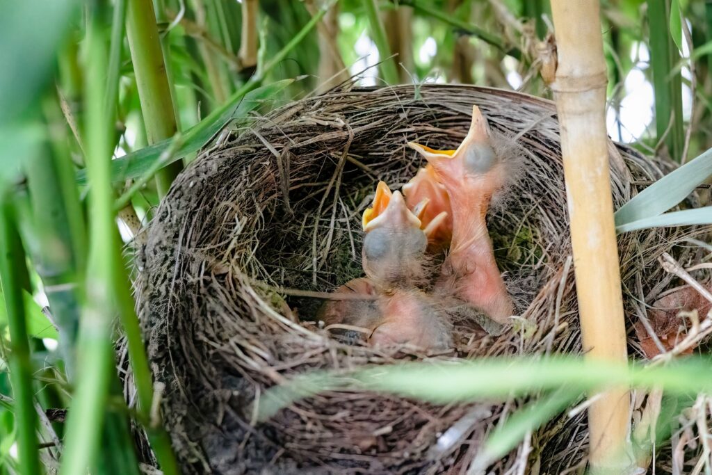
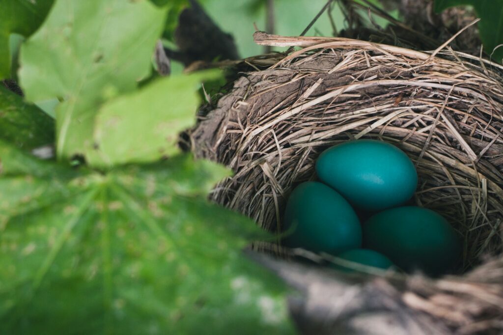
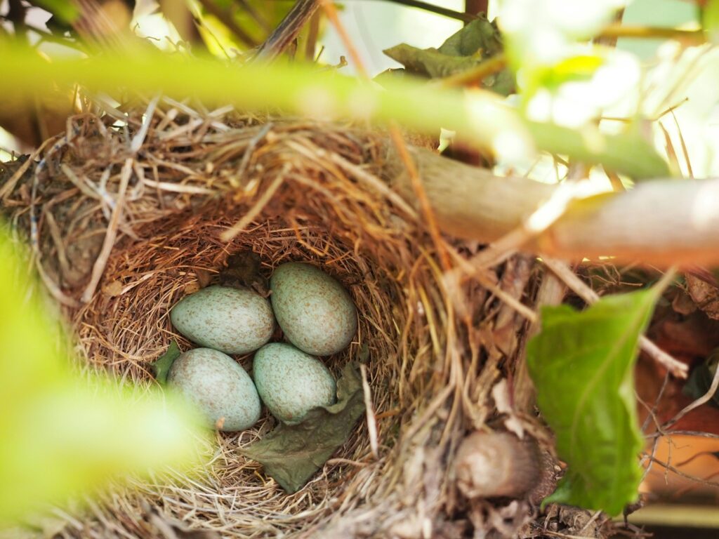
While the fundamental processes of avian embryonic development are similar across species, the timing and specific adaptations vary dramatically according to evolutionary history and ecological niche. Hummingbirds may complete incubation in as little as 14 days, while large birds of prey like eagles require up to 45 days, reflecting differences in metabolic rates and final body size. Precocial birds like chickens, ducks, and quail develop more extensively within the egg, hatching with open eyes, down feathers, and the ability to walk and feed themselves almost immediately. In contrast, altricial species like songbirds and parrots hatch in a relatively undeveloped state—naked, blind, and completely dependent on parental care. These differences are reflected in egg composition, with precocial species having yolks proportionally larger and richer in nutrients than altricial species. Even the rate of organ development varies, with species-specific priorities evident in the timing of different developmental milestones that reflect the survival needs of each type of bird after hatching. These variations represent evolutionary adaptations to different reproductive strategies, balancing the energy investment in egg production against the demands of post-hatching parental care.
Post-Hatching Transitions: From Embryo to Chick

The transition from embryo to independent chick involves remarkable physiological changes that begin during hatching and continue in the hours and days afterwards. Immediately after breaking free from the shell, the chick undergoes a critical transition from chorioallantoic respiration to full lung breathing, accompanied by changes in blood circulation as embryonic vessels regress and pulmonary circulation increases. The internalised yolk continues to provide nutrients as the digestive system matures and prepares to process external food. In many species, the chick’s body temperature regulation gradually develops, shifting from ectothermic (relying on external heat) to endothermic (generating internal body heat) as specialised metabolic tissues activate. The egg tooth, having served its essential purpose, typically falls off within days of hatching. Perhaps most visibly dramatic is the transformation of appearance as downy feathers dry and fluff out, giving the previously wet, exhausted hatchling the characteristic fluffy appearance we associate with baby birds. These coordinated physiological transitions represent the final stages of the developmental journey that began with a single fertilised cell and culminates in a new generation of birds ready to continue the cycle of life.
Conclusion
The journey from fertilised egg to hatching represents one of nature’s most remarkable developmental processes. Within the confined space of an egg, a single cell transforms into a complex organism with specialised systems perfectly adapted for avian life. This development follows a precise genetic program yet remains flexible enough to respond to environmental conditions. The self-contained nature of the egg has driven evolutionary adaptations unique to birds, from specialised extraembryonic membranes to the efficient utilisation of limited nutrients. As we’ve explored the intricate processes occurring inside a bird’s egg before hatching, we gain appreciation for the sophistication of nature’s design. Each crack in a hatching egg represents not just the beginning of a bird’s life, but the successful completion of an extraordinary developmental journey that has been refined over millions of years of evolution.